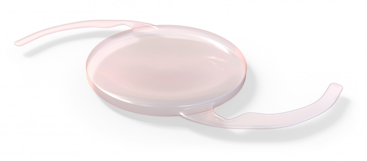
 Cataract surgery is often accompanied by an increase in growth of epithelial cells that line the back of the eye, leading to clouding of the lens — also known as posterior capsule opacification (PCO) — and decreased visual acuity.
A new study from the University of Florence in Italy and Poitiers University Hospital in France investigated how the design of a 360° square intraocular lens affects this increase in cell proliferation. The results, entitled “Square-Edge Intraocular Lenses and Epithelial Lens Cell Proliferation: Implications of Posterior Capsule Opacification in an In Vitro Model,” were published in BMC Opthalmology.
According to the American Academy of Opthalmology, the 360° square intraocular lens is the leading shape for intraocular lenses, with virtually all lens manufacturers using this design. “Without any doubt, the most important improvement to intraocular lenses with regard to PCO prevention is the incorporation of the square edge,” said Liliana Werner, MD, PhD, Assistant Professor of Ophthalmology at the University of Utah. “The square edge acts as a barrier, preventing the migration of lens epithelial cells from the equatorial region onto the posterior capsule.”
Yet even with this superior design, PCO is still clinically relevant, and Dr. Rita Mencucci, lead author of the study, indicated there is “a considerable interest in developing in vitro models allowing the clinician to understand the mechanisms of lens epithelial cell (LEC) proliferation.” Although other scientists have also seen the value of in vitro models to study PCO, their models are complex. Dr. Mencucci sought to simplify the model and use it to evaluate a one-piece and a three-piece intraocular lens, both from Abbott Medical Optics (AMO).
The research team grew human LECs on collagen membranes that were either in contact with an intraocular lens or plain. After six days, the researchers counted cells and evaluated their metabolic activity.
After analyzing the results, the researchers noted that, unsurprisingly, the greatest cell density was found under intraocular lenses. Considering the two different lenses, cell density was the same. “The present study shows that in our experimental conditions a 360° sharp optic edge acts as an effective barrier to LEC migration,” wrote Dr. Mencucci. This was an important piece of validation for the experiment, as it showed the model’s robust ability to mimic clinical findings.
“The main advantage of in vitro cell culture models is their acceptable reproducibility in the same experimental conditions,” wrote Dr. Mencucci, noting also that “it should be considered that this model, such as all the available in vitro models of PCO, is not able to fully reproduce the complex environment of the [eye].”


